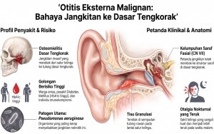
Bahagian 2: Lebih Daripada Sekadar Luka Biasa: Otitis Eksterna, Malignant Otitis Eksterna (MOE) Dan Perikondritis.
Jul 08, 2026
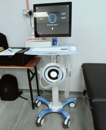
LOCATED IN THE SUBJECT PREPARATION ROOM AT THE BIOMEX BUILDING, AMDI., July 5 2025
Introduction
Osteoporosis and osteoporotic fractures are critical concerns for breast cancer survivors, significantly impacting their quality of life (Go et al., 2020). The non-ionizing radiation emitted by Radiofrequency Echographic Multi Spectrometry (REMS) makes it suitable for use in previously vulnerable groups, such as cancer survivors and enables more frequent monitoring of bone mineral density (BMD) (Fuggle et al., 2024). It is important to ensure adequate calcium intake among female cancer survivors (Zainordin et al., 2020) because cancer is a major risk factor for bone loss and fractures (Drake, 2013).
Problem Statement
Improved cancer survivorship means these patients may face chronic issues such as poor bone health (osteoporosis or osteopenia) and an increased risk of fractures (Wong and Chen, 2024) due to the long-term effects of cancer treatments such as chemotherapy, hormone therapy, or radiation (Rees-Punia et al., 2023). Despite the rising number of cancer survivors, awareness and implementation of bone health nutrition strategies remain limited. Without proper dietary knowledge and intervention, survivors are at higher risk for fractures, reduced mobility, and diminished quality of life. Therefore, targeted nutrition education is urgently needed to empower survivors with knowledge and skills to maintain bone health and prevent osteoporosis.
Objective
To evaluate the effectiveness of a nutrition education program in improving knowledge, dietary practices, and osteoporosis prevention behaviours among cancer survivors in Malaysia.
Methodology
Radiofrequency Echographic Multi Spectrometry (REMS)
Participants will lie on the provided bed. The avial site selection will be conducted to identify and prepare an appropriate anatomical location, ensuring optimal access, minimal discomfort, and reduced risk of complications. Following this, bone target visualization will be performed using REMS to accurately locate and identify the specific bone structure for osteoporosis diagnosis. The procedure for software-assisted ultrasound (US) acquisition will involve the use of specialized software to guide the capture, processing, and analysis of ultrasound images, ensuring precision and consistency in diagnostic imaging. Bone interface detection will be carried out through the calculation of Regions of Interest (ROIs), along with automatic signal and spectral analysis, to accurately identify and delineate the boundaries of bone structures. Finally, fragility score is a REMS measure of skeletal fragility (via bone microarchitecture and independent of BMD) at the spine and femoral neck and ranges from 0 (normal) to 100 (maximum fragility of the bone structure) (Fuggle et al., 2024). Figure 1 shows the REMS technology located in the subject preparation room at the Biomex Building, AMDI.
References