Dr Hazlin Hashim
Oncological and Radiological Sciences Cluster, Advanced Medical and Dental Institute,
Universiti Sains Malaysia, Bertam, 13200 Kepala Batas, Pulau Pinang.
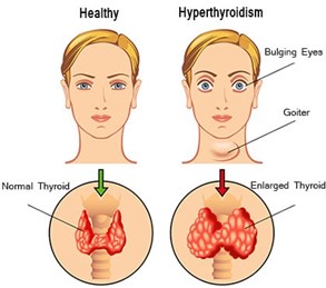
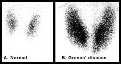
Hyperthyroidism (overactive thyroid) occurs when the thyroid gland produces too much of the thyroxine hormone. It can accelerate one body's metabolism, causing unintentional weight loss and a rapid or irregular heartbeat. Hyperthyroidism is a common endocrine disorder. The commonest aetiologies of hyperthyroidism are Graves’ disease and toxic multinodular goiter. The diagnosis of hyperthyroidism is usually made based on clinical symptoms and biochemical results. Ultrasonography of the neck and thyroid scintigraphy are used to evaluate the disease by identifying and characterising thyroid nodules as well as describing the anatomy of thyroid gland.
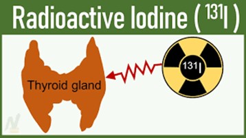
The initial treatment with anti-thyroid drugs, namely methimazole or propylthiouracil has been shown to lead to remission of Graves’ disease in approximately 60 % of cases with variation reported among studies involving the European population. However definite therapy with radioactive iodine (RAI) therapy and surgery is recommended in resistant and relapsed cases. Various methods of RAI dose determination have been in practice namely fixed, empirical and calculated dose.
Iodine-123 (123I) and Iodine-131 (131I) are used extensively in determining the percentage of thyroidal uptake in radioactive iodine uptake (RAIU) study. 123I is the ideal radiopharmaceutical for imaging and uptake measurement of thyroid gland because it has superior image quality and lower radiation exposure than 131I in addition to shorter overall procedure length. However, 123I is expensive and is not readily available. 99mTc-pertechnetate (99mTcO4) has been explored to be a suitable substitute in determining percentage thyroidal uptake. 99mTcO4 possesses similar characteristics to iodine but with better imaging qualities such as shorter imaging time, better image resolution, no beta radiation and lower overall radiation dose to the patient.
The advent of hybrid imaging such as single photon emission tomography / computed tomography (SPECT/CT) is able to quantitatively estimate the RAIU equivalent in thyroid gland using 99mTcO4 thyroid scintigraphy. Moreover, the utilisation of SPECT/CT has proved to be excellent in determining anatomical parameters such as volume measurement, an important parameter for dose calculation. However, the utilisation of fixed-dose RAI therapy has always been the preferred method for RAI therapy in many nuclear medicine centres in Malaysia.
Figure 1. (adapted from rmi.edu.pk/disease/hyperthyroidism)
Figure 2. Radioiodine therapy (I-131)
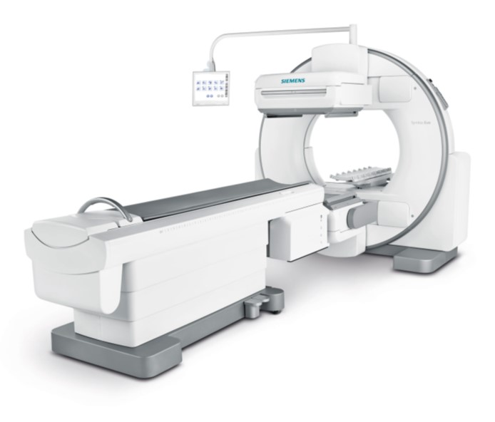
Figure 3. Gamma camera SPECT/CT (Siemens)
Figure 4. Thyroid scintigraphy (adapted from medbullets)
Penulis Artikel
Nama penulis:
Dr Hazlin binti Hashim
Afiliasi:
Kluster Sains Onkologi dan Radioterapi, IPPT USM
Bidang kepakaran:
Perubatan Nuklear